
Syringomyelia
Syringomyelia
What is Syringomyelia?
Syringomyelia, often referred to as SM, is a chronic disorder involving the spinal cord. The condition occurs when cerebrospinal fluid (normally found outside of the spinal cord and brain) enters the interior of the spinal cord, forming a cavity known as a syrinx.
This syrinx often expands and elongates over time, destroying the center of the spinal cord. As the nerve fibers inside the spinal cord are damaged, a wide variety of symptoms can occur, depending upon the size and location of the syrinx.
Causes of Syringomyelia
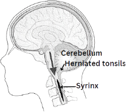 There are two major types of SM. In most cases it is related to a congenital malformation involving the hindbrain (cerebellum) called a Chiari I malformation. This malformation occurs during fetal development and is characterized by downward displacement of the lower part of the brain (cerebellar tonsils) beneath the foramen magnum, into the cervical spinal canal. This displacement blocks the normal flow of cerebrospinal fluid. When normal flow is obstructed, a syrinx can form in the spinal cord. Not all patients with Chiari malformations will develop a syrinx, however.
POST-TRAUMATIC SYRINGOMYELIA
Syringomyelia (SM) can also occur as a complication of trauma, meningitis, tumors, arachnoiditis, or a tethered spinal cord. In these cases, a fluid-filled cavity, or syrinx, forms in an area of the spinal cord affected by the underlying condition.
Post-traumatic syringomyelia (PTS) is an uncommon but potentially disabling complication of spinal cord injury (SCI). As more people survive spinal cord injuries, more cases of PTS are being diagnosed. A syrinx can develop years after the original injury. Scar tissue and changes in the spinal canal can disrupt the normal flow of cerebrospinal fluid (CSF), contributing to syrinx formation and expansion. Although cysts can form in more than half of people with SCI, only a small percentage progress to syringomyelia.
Activities that increase pressure, such as coughing, straining, heavy lifting, or breath-holding, may contribute to syrinx expansion. Symptoms and progression can vary widely, necessitating regular neurological and rehabilitation follow-up for detecting changes, managing symptoms, and helping prevent further neurological decline.
For more information, view these expert presentations from our annual conference:
- Syrinxes (Syringomyelia) Not Related to Chiari Malformations – Michael Levy, MD
- Primary Spinal Syringomyelia (Syrinx not Caused by Chiari) – Dr. Ulrich Batzdorf
- Syringomyelia in Chiari Patients – Konstantine Slavin, MD
DILATED CENTRAL CANAL VS SYRINGOMYELIA
A dilated central canal is generally a small, central enlargement of the spinal cord’s normal fluid-filled canal, while syringomyelia involves a larger, abnormal fluid-filled cavity (syrinx) within the spinal cord that can expand and potentially damage surrounding tissue.
Syringomyelia Symptoms
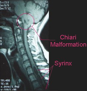 Symptoms of syringomyelia can vary considerably depending on the size and location of the syrinx and the areas of the spinal cord it affects. The syrinx often begins near the center of the spinal cord, where nerve fibers carrying pain and temperature information cross from one side to the other, resulting in pain and temperature sensation as the first senses affected. As the syrinx enlarges, it can affect additional nerve pathways, leading to weakness, muscle changes, balance and walking difficulties, and other neurological symptoms. Often, one side of the body may be affected more than the other. The mix of symptoms depends on which type of fibers and nerve cells are affected.
Symptoms often develop gradually over time or, in some cases, progress more quickly. Symptoms can sometimes appear or worsen suddenly following a fall or minor trauma. In children, scoliosis may be the only noticeable symptom. Some children with an atypical left-sided curve of the upper spine may undergo an imaging because of an atypical left-sided thoracic curve and discover a syrinx incidentally.
Symptoms can be subtle at first and may resemble those of other neurological conditions. Many individuals suffer from chronic pain, and some develop neuropathic pain syndromes, which can be difficult to treat. Syringomyelia can progress differently from person to person. New or worsening weakness, sensory changes, pain, or loss of function should be evaluated by a healthcare professional.
Tests & Treatment for Syringomyelia
CINE MRI
Syringomyelia is primarily diagnosed using magnetic resonance imaging (MRI), which can identify the location and size of a syrinx and show changes in the spinal cord or surrounding structures. A cine MRI may also be used to evaluate the flow of cerebrospinal fluid (CSF) and identify abnormalities or blockages in CSF circulation that may contribute to syrinx formation.
The first step after diagnosis is finding a neurosurgeon who is experienced in the treatment of SM. Finding a specialist is highly recommended. Surgery is the only viable treatment for SM, but not all patients will advance to the stage where surgery is needed. Evaluation of the condition is often difficult because SM can remain stationary for long periods of time, and in some cases progress rapidly.
Syringomyelia treatment is aimed at restoring the normal flow of spinal fluid. In persons with Chiari and SM, a procedure known as a posterior fossa decompression may be recommended. This procedure allows the cerebellar tonsils to move into a normal position, restoring the normal flow. After this procedure the syrinx will often reduce or resolve on its own. This can take months and may never completely collapse the syrinx. Some patients may require repeat surgeries. The most important consideration in deciding to undergo surgery is the severity of symptoms and overall quality of life issues.
In syrinxes associated with other causes, the surgical treatment is also aimed at restoring normal physiology and flow of spinal fluid. This might involve a bony spinal decompression, removing a tumor or scar tissue. In cases of tethered spinal cord, a surgical procedure is used to release the tension on the cord, allowing it to return to its normal position.
SYRINX SURGERY
A study of patients with CMI and SM examined how syrinxes and symptoms changed after craniocervical decompression and duraplasty. Nearly all patients experienced some improvement after surgery, and syrinxes generally became smaller over time. Syrinxes did not always disappear completely, and many patients continued to have symptoms, especially nerve pain and reduced sensation. The study found that patients with a smaller syrinx that remained after surgery had similar clinical outcomes to those whose syrinx resolved, suggesting that residual symptoms may reflect previous spinal cord injury rather than continued damage.
SYRINX SHUNTS
In rare cases, a shunting procedure may be necessary. This involves inserting a tiny tube into the syrinx which drains to a cavity outside of the spinal column. Shunting procedures are associated with certain risks and syrinx shunts frequently become clogged or dislodged. Repeated surgeries may be necessary.
Learn more about Syringomyelia surgery and treatment from these expert presentations:
- Understanding the How & Why of Surgery for Chiari in the Presence of Syringomyelia- Jeffrey Greenfield, MD
- Syrinx/Syringomyelia: When Should You Worry & Intervene? – Bermans Iskandar, MD
- Syringomyelia Update – John Heiss, MD
LIVING WITH SYRINGOMYELIA
Living with syringomyelia can mean managing chronic pain, sensory changes, and other symptoms that may affect daily life. Finding ways to manage symptoms, maintain overall wellness, and stay connected to supportive resources can be an important part of living with CM/SM.
Explore ASAP’s Wellness Resources for Patients for information on pain management, exercise, sleep, stress, heat intolerance, and coping with syringomyelia and Chiari.
Should CM/SM Patients Undergo a Spinal Tap?
A lumbar puncture (spinal tap) is a procedure in which spinal fluid is removed from the subarachnoid space in the lumbar region of the spine. It is often ordered to check for infection, rule out certain diseases, or to detect and/or treat elevations in spinal fluid pressure.
Individuals often ask if a lumbar puncture is contraindicated in people diagnosed with Chiari malformation and/or syringomyelia. Some research shows that having CM may increase the risks associated with this procedure. Some authors have reported a worsening of symptoms or catastrophic results following the procedure, but these are considered very rare. In those patients who have been successfully surgically treated, these concerns are even less of an issue.
Ensure the ordering physician is aware of your diagnosis and discuss all potential benefits and risks of the procedure. If the situation is not an emergency, it is a good idea to discuss the procedure with the neurosurgeon. Your medical team can determine whether the potential benefits outweigh the risks. It is important to remember that some conditions such as meningitis can be fatal if not diagnosed and treated properly.