by Arnold H. Menezes, MD
The following article is a transcribed presentation from the 2004 ASAP Conference.
Abstract
The clinically symptomatic child with the hindbrain herniation syndrome (Chiari I) can differ significantly from the adult patient. A prospective analysis of these children has brought an understanding of the various presentations.
The primary symptoms below age 5 have been failure to thrive secondary to repeated aspirations, gastroesophageal reflux and headaches. Between ages 3-10 years, headaches and scoliosis were the main symptoms together with neurological abnormalities secondary to the cerebellar tonsillar impaction and syringohydromyelia.
The factors affecting treatment have been clinical symptoms/signs, posterior fossa volume, craniovertebral junction abnormalities and instability. Taken together, these substrates provide for successful outcome.
Transcribed Presentation Notes


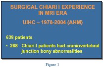
Ive been at the University of Iowa since 1969 [see Figure 1] and my interest in terms of keeping a detailed log started out fairly early. We have a prospective data base and it looked at nearly 600 surgical patients, because of my interest in abnormalities of the skull base, bony abnormalities, about 2/5 of them, nearly half of them, were patients who had problems with skull base abnormalities. But when we focused on children below the age of 6 years, [see Figure 2] we thought that our reason for doing this would be, how did this defer in terms of presentation from the adults or anybody above that age? And why did we pick the age of 6? Because it begins to be statistically significantly. We wanted to achieve an early diagnosis, and more importantly, we need to educate medical practioners, such as pediatricians, family practioners, general practice, neurosurgeons too, and I think that that was something we needed to achieve. Once we came to some conclusions, we thought that our best bet was to have special meetings, instead of going ahead and presenting this at neurosurgical national meetings, which is what we do, but also to educate the pediatricians. The Journal of Pediatrics has a circulation of over 300,000 and its translated into 4 languages and goes out world wide.
