Diagnostic Studies: the MRI
Magnetic resonance imaging (MRI) has revolutionized neurological diagnosis. It gives detailed pictures of the brain, the spinal cord, and the surrounding tissues. When people without neurological symptoms undergo an MRI of the brain, the tonsils are found to be an average of 3 mm above the foramen magnum.
 The figure above shows two MRI scans. The scan on the left is normal and shows cisterna magna. The tonsils are above the foramen magnum. The MRI on the right is that of a patient with the Chiari I malformation. The tonsils hang down below the foramen magnum and crowd out the cisterna magna, which is no longer visible. In this case, there is also a kink at the junction between the brain stem and spinal cord.
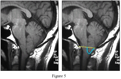
 The Chiari I malformation is present when the tonsils herniate more than 3 or 5 mm below the foramen magnum. [see Figure 5] The measurement is taken by drawing a line from the end of the clivus bone (called the basion) to the end of the supraocciput (called the ophistheon). Another important finding is the amount of crowding at the foramen magnum.

