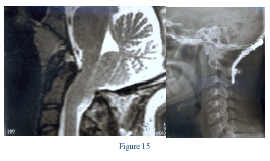
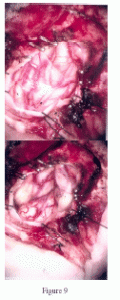
 This is through the microscope, [see Figure 9] shows you the tonsils are down, this is the left side, the right side, and its squeezed down. We shrink them, make them go up or move them so that the opening through the cavity in the brain, the 4th ventricle is still exposed, [see Figure 10] and then we finish it off making sure that everything is wide open and satisfactory and keep it in place. And the analogy I give my patients and parents, if I have a waist of 38 and I have a belt of 34 Im in trouble, so either I get a new belt or I splice a new patch [see Figure 11] which means that I have to increase it to 40 so that I can breath which is exactly what she is doing, which is called a dural graft.
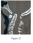
 So thats one side of it. [see Figure 12] But what about this patient such as this young man who is 6 years old? He has an abnormality; this is the space here through which the brain should come down into the spine. Well, half of that is taken up with this bone that is projecting into it, thats called basilar, and there is an abnormality here because the second bone is stuck to the third front and back and its pushed up. This should have been right here. Can we avoid 2 operations? Thats the important thing that means going from in front -big procedure, through the mouth; taking this out, going from behind, try to make it only one operation. So what we do is we try and get this patient into traction and
pull it down. The 3D CT, this is just to show you the hologram of 3D rendition of the problem. It shows you that things have abnormal alignment. Youre looking at the spine and skull just as though youre holding it in your hand from in front. In traction we were able
to pull that bone which was up here down so that the space here is correct and we could get by with an operation only from behind.
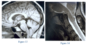
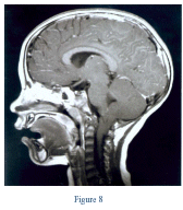
This is a child who stopped dancing because every time she did a cartwheel or a flip she got into trouble. This MRI shows that shes got this portion of her cerebellum hanging down and the area looks pretty abnormal. [see Figure 13] This should have been a nice curve down here, instead its dented backwards. So instead of having a convexity forward shes got a concavity– that means weve got a problem in front. And this is what we talk about flexion/extension MRIs. We get an MRI, a control study and we are seeing that this dent here, [see Figure 14] focus on this, that dent needs to go, either with an operation, or position, or traction. In addition weve found that if this dent has this blood vessel over it and bangs against it, 25% of these children come in with headaches, and with the older patients its called migraines. Why does that happen? Because that blood vessel gets banged against, you stop it, put them in traction or fuse them, and it is relieved.
