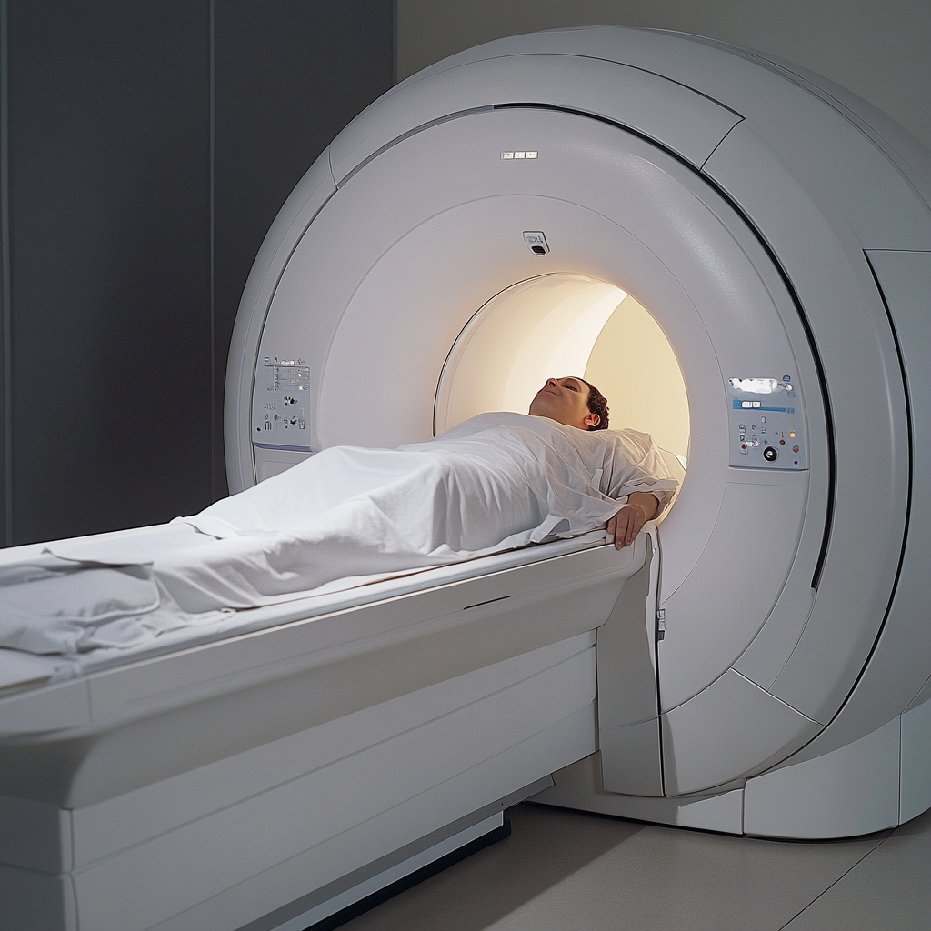
Cine MRI
Cine MRI
Cine MRI – A specialized type of magnetic resonance imaging
A Cine MRI (cine phase-contrast MRI), pronounced “sin-ee” is a specialized type of magnetic resonance imaging that captures moving images, allowing doctors to observe the flow of cerebrospinal fluid (CSF) and other dynamic processes in real-time. “Cine” is derived from the word “cinema,” reflecting its capability to create a series of images that show motion, similar to a movie.
This MRI is taken the same way as a traditional MRI, with the addition of either a wristband or EKG leads on the patient’s chest to measure the heart rate. Each time your heart beats, the cerebrospinal fluid is forced out of your brain, down toward the spine in response to the flow of blood that enters the brain with each beat. The MRI machine is equipped with an additional software package that allows the images to be put together, showing the flow of the cerebrospinal fluid (CSF) as it is moving.

Procedure:
- Preparation: The patient is positioned in the MRI machine, similar to a standard MRI procedure. They may need to hold their breath or follow specific instructions to ensure clear images.
- Imaging: The MRI scanner uses magnetic fields and radio waves to create detailed images. For cine MRI, the scanner takes multiple images over a period, creating a sequence that shows movement.
- Analysis: Radiologists and specialists analyze the cine MRI images to determine the amount of fluid that is moving and compare that with normal subjects.
Cine MRI Usage in Chiari Malformation Diagnosis:
- CSF Flow Assessment: Cine MRI is particularly useful in Chiari malformation for evaluating the flow of CSF at the foramen magnum (the opening at the base of the skull). It helps determine if the herniated cerebellar tonsils are blocking CSF flow, which could result in syringomyelia or hydrocephalus. The presence of these additional symptoms can guide treatment decisions.
- Chiari 0: Cine MRI’s can be useful in cases of “borderline” Chiari malformations or when the question of whether or not decompression is needed is not readily answered using a traditional MRI.
- Pre- and Post-Surgery: It can be used both before and after decompression surgery to evaluate the effectiveness of the procedure in restoring normal CSF flow.
Cine MRI scans are not as widely available as standard MRI scans. This limited availability can be due to the specialized nature of the equipment and expertise required to perform and interpret cine MRI studies. The practical use of cine MRI in clinical practice is still a topic of debate among physicians. Some clinicians find it valuable for diagnosing and managing conditions like Chiari malformation and hydrocephalus, while others question its necessity and cost-effectiveness compared to other diagnostic methods.
Related Articles
Related Videos
Craniocervical Anatomy in Black & White – Carina Yang, MD
The Fundamentals of Chiari & Syringomyelia from a Neuroradiologist’s Perspective – Carina Yang, MD
Advanced Imaging Technologies & AI for Chiari Malformation & Beyond – Moss Zhao, DPhil
A Glimpse Inside the MRI of Chiari, Syringomyelia & Related Disorders – Carina Yang, MD
Engineering-based Diagnostics for Assessment of Chiari Malformation – Gwen Williams, MD

