Research
Research
The Crucial Role of Research in Advancing Knowledge and Innovation
Research is essential in the search for cures, as it provides the foundation for understanding complex biological processes, identifying causes of illness, and developing effective treatments. Clinical trials, laboratory experiments, and data analysis are all key components that help translate scientific discoveries into real-world medical breakthroughs. Without research, advancements in medicine would stagnate, making it impossible to find cures for both common and rare diseases, ultimately saving lives and improving global health outcomes.

Current Research
Project Title: Validating an Analytic Model for Cerebrospinal Fluid Flow Across the Foramen Magnum in Children with Chiari I Malformation with Syringomyelia: A Study of Patients Before and After Surgery
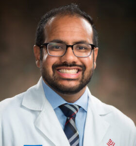
 Grant Recipient: Vijay M Ravindra, MD, MSPH
Institution: University of Utah/Primary Children’s Hospital
Authors: Vijay M. Ravindra, MD, MSPH, Guillermo L. Nozaleda, BS, Douglas L. Brockmeyer, MD, Antonio
L. Sánchez, PhD, John Rampton, MD, Mark Quigley, PhD
Date: September 1, 2024 through August 31, 2025
Grant: $53,082
Summary of Research:
In a prospective study of children with Chiari malformation with Syringomyelia, they aim to numerically model the velocity of CSF flow at the craniocervical junction using mathematical equations and compare the findings against traditional MRI measurements of CSF flow.
The proposed study aims to develop an initial analytic understanding of this complex region, which represents a significant knowledge gap. We anticipate these findings will serve as pilot data and a stepping stone to understanding fluid-flow implications of the disease process, with hopes of tailoring treatment for children with syringomyelia based on the information gathered.
Project Title: Tonsillar Manipulation During Chiari I Malformation Surgery

Institution: Children’s National
Date: April 1, 2024 through December 31, 2024
Grant: $17,722
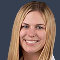
This funding is provided to Kelsi Chesney, MD, recipient of the Timothy M. George Fellowship Award. Dr. Chesney writes, “I am particularly drawn to the multi-institutional collaboration that this project plans for. I firmly believe that the future of medicine lies in collective efforts of diverse experts working together to solve complex problems.”
Summary of Research:
Current literature on the surgical technique for Chiari malformation (CM) consists of case series with variable cohort sizes and follow-up times, yielding disparate results and emphasizing the intricacies of clinical decision-making when dealing with CM patients. Multiple series have demonstrated that tonsillar manipulation is associated with increased postoperative complication risk, such as pseudomeningocele, hygroma formation and meningitis, as well as higher rates of treatment failure. However, a recent survey of international experts in the field of CM shows that the most experienced surgeons agree that more invasive techniques are the most effective.
While posterior cranial fossa decompression remains the standard of care for symptomatic CM, the extent of intradural work, particularly tonsillar manipulation, remains a contentious issue. The goal of this fellowship project will be to investigate the extent of tonsillar manipulation during surgical treatment of CM as it relates to multiple outcome parameters through extensive data collection across multiple institutions.
Project Title: Assessing the Physical Impact of Chiari in Adults
Grant Recipient: Philip A Allen, PhD
Institution: University of Akron
Date: November 2023
Grant: $33,500
Summary of Research:
While the presenting symptoms of Chiari I in adults are well documented, the long-term physical impact and manifestations are not. This project will, for the first time, characterize and quantify the physical impact of Chiari on a large group of adult patients. This will be accomplished through a web survey comprised of widely used assessments covering topics such as headaches, sleep, neck disability, upper/lower body function, balance, and more. In addition, a pilot project will be undertaken to collect in-depth information on a small group of patients through in-person evaluation and biomechanical testing by a licensed physical therapist. The goal of the pilot study is to identify underlying causes of functional limitations to inform the development of future interventions that can improve quality of life.
Key findings and Next Steps
Project Title: Medicinal Marijuana for the Treatment of Pain in Patients with Chiari Malformation
Grant Recipient: Erol Veznedaroglu, MD
Institution: Global Neurosciences
Date: June 2021
Grant: $59,000
Summary of Research:
Although anecdotal evidence of the pain-relieving effects of medicinal marijuana (MM) is abundant, clinical data does not exist for pain management with MM in adult patients with Chiari malformation (CM). The study co-investigated by Dr Erol Veznedaroglu, Global Neurosciences Institute and Dr Ruth Perry, Cannabis Education and Research Institute, will survey ASAP members with CM to determine their use, frequency, dosage and strain of MM. Participants will be evaluated to determine if they are getting optimal pain relief from their MM strain. The study should provide evidence for an optimal strain of MM to treat pain associated with CM.
A ’patient experience registry’, based on treatment and patient experience data, collected from consenting qualifying patients will be used to analyze the efficacy and side effects of various strains of marijuana in the treatment of CM related pain, and provide actionable information to physicians, patients caregivers, alternative treatment centers and others in support of the effective use of medical marijuana in this patient population.
Project Title: DNA Methylation in Familial Chiari I Malformation
Grant Recipients: Bermans Iskandar, MD and Reid Alisch, PhD
Institution: University of Wisconsin
Date: May 2021
Grant: $99,999.41
Summary of Research:
While twin and family studies suggest that Chiari I malformations are genetically inherited, genetic mutations have not been identified as a consistent cause of theses malformations. Epigenetic modifications contribute to heritable conditions that can be influenced by environmental factors without a change in the DNA sequence. Thus, it is believed that alterations in epigenetic modifications cause inherited forms of Chiari I that lack an obvious genetic predisposition but have perceptible lines to environmental conditions.
With saliva obtained from patients with familial Chiari I and unaffected controls, they will investigate the role of epigenetics, specifically DNA methylation, in Chiari I families. Findings from this research will provide critical molecular insights in the heritable basis of these malformations and guide future research directions.
Completed Research
Project Title: Histological Investigation of the Posterior Atlanto-Occipital Membrane in Pediatric Patients with Chiari I Malformation
Grant Recipient: Vijay M Ravindra, MD and Douglas Brockmeyer, MD
Institution: University of Utah
Date: July 2021
Grant: $19,982.00
Read more about the study here
Summary of Research:
Prospective study of children with Chiari I Malformation, we aim to histologically examine the posterior atlantooccipital membrane (PAOM) after its removal in surgery and compare the findings with those of controls. We hope this study will serve to develop a more complete understanding of an important ligamentous structure and anticipate these results will serve as pilot data to further study the importance of the PAOM as it relates to outcomes in CM-I, specifically when comparing posterior fossa decompression with and without duraplasty. Future therapeutics targeting this membrane may represent an addition avenue for further research. We hypothesize there are structural differences in PAOM in children with CM-I and controls that contributes to disrupted flow of CSF in the foramen magnum.
The study will be executed by Vijay M Ravindra, who is a pediatric neurosurgeon at the Naval Medical Center San Diego with a faculty appointment at the University of Utah, under the supervision of Dr Douglas Brockmeyer at the University of Utah/Primary Children’s Hospital. All patients will be enrolled at the University of Utah/Primary Children’s Hospital.
Project Title: Quantifying gait and postural control in Chiari patients 2020
Principal Investigator: Brian Davis, PhD
Institution: Akron Children’s Hospital
Date: July 2020
Grant: $49,896
Summary of Research:
Chiari Malformation (CM) is a disorder at the junction of the skull and spine. It is associated with protrusion of the base of the brain into the top of the spine, thus inhibiting flow of cerebrospinal fluid. This results in pressure buildup, causing the headaches, dizziness, difficulty swallowing, muscle weakness, and loss of neuromuscular coordination. The last of these symptoms is the focus of the proposed research.
Specifically, this study aims to quantify postural stability in pediatric patients with CM. Although loss of stability and impaired walking are frequent symptoms, no previous studies have examined either of these deficits in CM patients. Understanding neuromuscular control will provide further insights to neurologists, neurosurgeons planning surgical intervention and to therapists designing pain reduction and restorative balance techniques.
Between 2012 and 2019, 192 patients were treated at Akron Children’s Hospital (ACH) with a diagnosis of Arnold-Chiari syndrome without spina bifida or hydrocephalus. The proposed project will recruit 20 of these pediatric patients with SM and 20 age and sex matched controls and measure neuromuscular coordination using two simple tests including (i) the ability to stand upright and motionless for 30 second durations and (ii) the ability to perform simple waling trials across an instrumented walkway. Data will be collected at baseline and again at six months to document (i) differences in age-matched control subjects and (ii) changes over time. These data will serve as baseline for all future studies that examine neuromuscular coordination in CM patients and help inform clinical practice using objective measures of symptoms and their management.
The team that has been assembled for this research project includes Dr Gayathri Sreedher, an experience pediatric neuro-radiologist based at ACH (the 7th largest pediatric hospital in the USA), Dr Brian Davis, former President of the International Society of Biomechanics and co-author of a textbook on human gait, and Dr Doug Wajda from Cleveland State University and an expert on fall risk assessment.
Quality Indicators in Pediatric Chiari I Grant
Recipient: Gerald Grant, MD
Institution: Stanford University
Date: May 1, 2020
Grant: 102,497.39
Summary of Research:
Chiari I malformation is an anatomical abnormality characterized by a number of anatomic and radiologic findings. Classically, children can present with headache, long tract signs (such as loss of sensation, position sense, weakness, spasticity, or incontinence), bulbar symptoms (such as difficulty swallowing, dysarthria, or other dysfunction of the lower cranial nerves), or other neurologic impairments. There are also impairments across physical, psychological, and social domains in patients with Chiari I. Surgical decompression is a common implementation of treatment. Our goal is to establish the “standard of care” for the surgical management of Chiari I malformation. Despite quality of life measures to assess the surgical outcomes of Chiari decompression, the natural history is relatively unknown. We will use an evidence-based approach to establish Chiari indicators using a Collaborative Health Outcomes Information Registry (Peds-CHOIR) to inform clinical and surgical decision-making. We have reviewed the literature in an unbiased and systematic fashion to identify variables that can either predict whether a patient will benefit from surgery or whether a surgery has been “successful.” We will apply these variables prospectively before and after surgery.
Amplified Imaging of the Pediatric Brain
Grant Recipient: Michael Moseley, MD
Institution: Leland Stanford Junior University
Date: May 2020
Grant: $90,728.00
Summary: The research project will create and test a new way of illustrating and mapping small brain dynamics using a novel MRI method, “amplified Magnetic Resonance Imaging” (aMRI). This method takes conventional series of MR images of the pediatric brain and processes these into a ‘cine’ or movie loop that can amplify intrinsic motions from the pulsing arteries on the surrounding brain to detect a variety of subtle brain and spinal cord dynamic abnormalities. We anticipate that the application of amplified movies of the brain will become an important tool in pediatric imaging and offer unique visualizations of altered or abnormal structure and function in potentially any tissue from any modality. We expect that this new tissue diagnostic mechanism will rapidly advance precision health in children.
Comparative Analysis of In Vivo and In Vitro CSF flow Studies in Chiari Malformation
Grant Recipients: Bryn Martin/Gwen Williams, MDs
Institution: University of Idaho
Date: January 2019
Grant: $47,084.00
Long-term outcomes for children with incidentally discovered Chiari malformation type 1: what is the clinical significance?
Grant Recipient: Robert F Keathing, MD
Team: Laurence Davidson, Tiffany N Phan, John S Myseros, Suresh N Magge, Chima Oluigbo, Carlos E Sanchez, Robert F Keating
Date: November 2020
Objectives
Chiari malformation type 1 (CM1) is an increasingly common incidental finding on magnetic resonance imaging (MRI). The proportion of children with an incidentally discovered CM1 who upon further evaluation require operative intervention for previously unrecognized signs and symptoms of neurological compromise or significant radiographic findings (syringomyelia) is unclear. An extensive long-term single-institution patient series was evaluated to better clarify the likelihood of surgery in patients who present with an incidentally discovered CM1.
Long-term outcomes of posterior fossa decompression for Chiari malformation type 1: which patients are most prone to failure?
Grant Recipients: Robert F. Keating, MD
Date: July 2021
Objectives
The role of an osseous-only posterior fossa decompression (PFD) for Chiari malformation type 1 (CM1) remains controversial. We reviewed long-term outcomes for patients with CM1 undergoing a PFD to evaluate if there was any difference for failure when compared to patients undergoing a PFD with duraplasty (PFDD).
Effects of demographic and socioeconomic factos in diagnosis and treatment of CMI
Grant Recipient: Dave Limbrick, MD
Institution: Washington University in St. Louis
Date: March 2017
Grant: $29,000.00
Project title: Postoperative Syringomyelia Resolution in Canine Chiari Like Malformation
Grant Recipient: Lauren R Talarico, BS, DVM, CACVIM
Institution: VCA SouthPaws Veterinary Specialty and Emergency Center
Date: December 2015
Grant: $57,000.00
Summary of Research: Malformation of the caudal aspect of the skull, similar to Chiari type I disorder of people, has been documented in many breeds of dogs. Even though approximately 95% of all Cavalier King Charles Spaniels are affected by Chiari-like malformation (CLM) other breeds of dogs such as Yorkshire Terriers and Chihuahuas are also commonly affected.
Aims:
1. To determine objective measurements of syrinx size pre and post-operatively and how changes in the size of the syrinx differs between persistently symptomatic and clinically improved postoperative patients.
2. To correlate the above findings with published human data to determine if cross species differences exist and to look for etiologic clues for this disease in both populations, particularly looking at the variable presentation of Chiari in the human population. This may also help to define which patients are at risk due to the lack of posterior fossa volume versus an obstruction of CSF flow at the outlet.
Project title: Quantifying CSF Flow Dynamics Using Real-time MRI
Grant Recipient: Karim Sabra, PhD
Institution: Georgia Institute of Technology
Date: September 2015
Grant: $50,000.00
Summary of Research: This project aims 1) to quantitatively measure in real-time and non-invasively the cerebrospinal fluid (CSF) flow dynamics using a novel magnetic resonance imaging (MRI) technique in order to investigate the effects of respiration and transient events, such as coughing and sneezing, on SCF flow along the spine, and 2) to determine the applicability of this MRI method to improve the diagnosis of craniospinal disorders and better understand the role CSF flow plays in the pain symptoms experienced by Chiari malformation and syringomyelia patients.
ASAP Chiari Research Fellowship
Assessment of 4D PC MRI…Cerebrospinal fluid motion
Grant Recipient: Bryn Martin
Institution: University of Akron
Date: March 2013
Grant $53,568
Developing a preoperative Chiari severity index
Grant Recipient: Dave Limbrick
Institution: Washington University in St. Louis
Date: May 2013
Grant: $25,000.00
Dr David Limbrick is a pediatric neurosurgeon at Washington University and St Louis Children’s Hospital. He will be responsible for managing and coordinating the research. He will supervise patient recruitment, IRB submission and compliance. Dr. Limbrick’s project title is Developing a Preoperative Chiari Severity Index to Stratify Prospective Clinical Studies for Chiari type I Malformation.
Due to the dearth of rigorously-acquired, prospective clinical data, the optimal treatment of Chiari malformation I (CM-i) remains one of the most controversial topics in pediatric neurosurgery. While recent research efforts have yielded scales for assessing post-surgical outcomes, effective metrics for stratification of patients at the time of initial presentation (before any surgical intervention) are lacking. The development of such a metric is crucial to designing and conducting clinical trials to assess differential efficacy of the various surgical approaches for CM-I.
The current project will use conjunctive consolidation methodologies to create the Chiari Severity Index (CSI), a multi-dimensional composite index that classifies the severity of clinical and radiographic findings for children presenting with CM-I. After validation in an ongoing multi-institutional study of CM-I and syringomyelia (the Park-Reeves Syringomyelia Research Consortium), the CSI will serve two critical roles:
1) Providing meaningful prognostic information to anticipate clinical outcomes in CM-I,
2) Stratifying patients for prospective clinical trials evaluating the differential efficacy of major CM-I treatment approaches.
An urgent need to identify optimal treatment approaches for CM-I is clearly evident. The CSI represents the first and most logical step in this important process.
Is CSF Flow the “Holy Grail” to determining Clinic-pathological Significance of Chiari Malformation?
Grant Recipient: Mark Quigley, MD
Institution: Children’s National
Date: May 2013
Grant: $21,000.00
Analysis of Posterior Fossa Volumes
Grant Recipient: Alan Sui, MD
Grant Institution: Children’s National
Date: May 2015
Grant $12,025.00
Disease Progression of Pediatric Patients …CMI
Grant Recipient: Kerri Thorn, MD
Institution: Children’s National
Date August 2009
Grant: $8,500.00
Purpose of the study: Some patients are found to have Chiari I malformation after they have an MRI scan of the head or neck to evaluate for another condition. The finding of Chiari I Malformation on the MRI study is considered to be “incidental Chiari I malformation,” which requires no treatment, if symptoms are thought to be unrelated to Chiari I Malformation. The fate of patients with incidental Chiari I malformation is uncertain. Research is required to answer these important questions: 1) What percentage of patients with incidental Chiari I malformation will later develop symptoms and require treatment for Chiari I malformation? 2) Can specific findings on MRI scans predict who will develop symptomatic Chiari I malformation? 3) Are there data to support routine follow-up MRI scanning in these patients?
Researchers at Children’s National Medical Center, Washington, DC, are performing a ten-year retrospective review of their database of patients with “incidental Chiari I malformation” and “symptomatic Chiari I malformation.” They will evaluate whether patients with the diagnosis of incidental Chiari I malformation developed symptomatic Chiari I malformation during that time period. The study will also compare symptoms and MRI findings of patients with incidental Chiari I malformation with those of patients with symptomatic Chiari I malformation and evaluate for factors that predict development of symptomatic Chiari I malformation. The project has already received Investigational Review Board (IRB) approval. Kerri Thorn, PA-C., is the Principal Investigator on the project and her Associated Investigators include a pediatric neurosurgeon and a physician assistant. An ASAP Grant has been awarded and research will start immediatel
Cognitive Function …before and after surgery
Grant Recipient: John Oro
Institution: Friends Research Institute
Date: October 2008
Grant $24,020.00
ASAP Chiari Research Fellowship /Washington University in St. Louis
Principal Investigators: Spiros Blackburn MD
Institution: Congress of Neurological Surgeons
Date: July 2008
Grant $10,000.00
Outcomes in Patients Undergoing Surgical Intervention for Chiari Type I Malformation with Syringomyelia
Grant Recipients: Bermans J. Iskandar, MD
Director of the Pediatric Neurosurgery Program, University of Wisconsin/Madison.
Tim M. George, MD, University of Texas at Austin
Date: May 2007
Grant: $220,000
Fellowship
Grant Recipient: Sandi Lam, MD
Institution: Congress of Neurological Surgeons
Dates: March, 2007
Grant: $10,000
Fellowship
Grant Recipient: Matthew McGirt, MD
Institution: Congress of Neurological Surgeons
Dates: March 2007
Grant: $10,000
Fellowship
Grant Recipient: Andrew Jea, MD
Institution: Congress of Neurological Surgeons
Dates: January 2006
Grant: $10,000
Investigation on the pathogenesis of post-traumatic syringomyelia (PTS): the roles of central canal occlusion and focal arachnoiditis on the contused and intact rat spinal cord
Grant Recipient: Christopher Shields, MD, Professor and Chairman
Institution: University of Louisville School of Medicine, Louisville, KY
Dates: September 2005
Grant: $50,000
Summary of Research:
Syringomyelia is a major complication following spinal cord injury. Its occurrence causes pain and additional neurological impairments. Treatment of syringomyelia following spinal cord injury is limited. The absence of an animal model of post-traumatic syringomyelia (PTS) limits our ability to develop appropriate treatments for this condition. Using a rat, we believe that a model of PTS can be created by surgically induced focal arachnoiditis in the cervical area. We believe that the creation of PTS will be particularly fruitful following an associated spinal cord injury. We plan to surgically create a model that will cause post-traumatic syringomyelia. Animals will be monitored for one year following the surgery by sequential cervical MRI scans, and electrophysiological techniques that monitor descending motor pathways and ascending sensory pathways. Moreover, the rats will be sacrificed to assess the morphological changes on year after surgery. We postulate that if PTS does develop, there will be significant behavioral, electrophysiological, imagine, and morphological changes. If we are successful in creating such a model, this will allow the development of a series of innovative therapies for the management of PTS.
Importance of the Mechanical Forces in the Pathogenesis of Syringomyelia
Grant Recipient: Francis Loth, PhD, Department of Mechanical and Industrial Engineering
Institution: University of Illinois at Chicago
Dates: September, 2005
Grant: $50,000
Summary of Research:
A simplified in vitro model of the spinal canal, based on in vivo magnetic resonance imaging (MRI), was used to examine the hydrodynamics of the human spinal cord and subarachnoid space (SAS) with syringomyelia. In vivo MRI measurements of SAS geometry and cerebrospinal fluid (CSF) velocity were acquired in a patient with syringomyelia and used to aid in the in vitro model design and experiment. The in vitro model contained a fluid-filled coaxial elastic tube to represent a syrinx. A computer controlled pulsatile pump was used to subject the in vitro model to a CSF flow waveform representative of that measured in vivo. Transducers measured unsteady pressure both in the SAS and intra-syrinx at four axial locations in the model.
MRI results indicated that the peak-to-peak amplitude of the SAS flow waveform in vivo was approximately ten fold that of the syrinx and in phase. The in vitro flow waveform approximated the in vivo peak-to-peak magnitude. Peak-to-peak in vitro pressure variation in both the SAS and syrinx was approximately 6 mmHg. Syrinx pressure waveform lead the SAS pressure waveform by approximately 40ms. Syrinx pressure was found to be less than the SAS for ~200 ms during the 860 ms flow cycle. Unsteady pulse wave velocity (PWV) in the syrinx was computed to be a maximum of ~25 m/s. Spinal cord wall motion was found to be non-axisymmetric with a maximum displacement of ~140 m, which is below the resolution limit of MRI.
Agreement between in vivo and in vitro MR measurements demonstrated that the hydrodynamics present in the fluid filled coaxial elastic tube system are similar to those present in syringomyelia. Overall, the in vitro study of the unsteady pressure and flow environment within the syrinx and SAS, provides insight into the complex biomechanical forces present in syringomyelia.
We propose to continue experimentation using this in vitro model of syringomyelia in order to determine the influence of various configuration changes on the hydrodynamic environment within the syrinx and SAS. Our previous research demonstrated a phase shift between the syrinx and SAS pressure that could provide a mechanism for syrinx progression. Thus, the proposed work would examine this potential mechanism under various configurations thought to cause syringomyelia. These configuration changes would represent conditions such as coughing, Chiari malformation, flow obstruction due to vertebra misalignment, changes in atmospheric pressure, and spinal cord tension. We will focus on obtaining measurements that provide a more complete understanding of the role of hydrodynamic forces in syringomyelia pathogenesis. Finally, we will continue research to develop novel MRI techniques that will provide detailed information about patient geometry to better assess syringomyelia and Chiari malformation severity.
Fellowship
Grant Recipient: Richard Reid, MD
Institution: Congress of Neurological Surgeons
Dates: January 2005
Grant: $10,000
Pre-clinical Development of GABA Cell Therapy For Chronic Pain After Spinal Cord Injury (year 3)
Grant Recipient: Mary Eaton, PhD, Assistant Professor of Neurological Surgery
Institution: University of Miami
Dates: October 2004
Grant Amount: $50,000
Summary of Research
Backed by the resources of the Miami Project To Cure Paralysis, Dr. Eaton’s research focused on developing cellular ‘minipumps’ injected near pain-processing areas of the spinal cord to both alleviate and potentially eliminate chronic, neuropathic pain. Dr. Eaton developed cells that secreted (or pumped) GABA, a naturally occurring neurotransmitter in the spinal cord that inhibits pain, and injected the cells into rats with spinal cord injuries. Dr. Eaton’s research suggested that in a severe spinal cord injury, not enough GABA is produced to cope with the injury, and chronic pain ensues. Dr. Eaton hypothesized that inadequate GABA can be compensated for by injecting a ‘minipump’ of cells that secrete GABA near the injury. After developing the GABA-secreting cells, Dr. Eaton studied how well they produced GABA in rats with spinal cord injuries and whether the increased GABA reduced indications of neuropathic pain.
Dr. Eaton’s research showed that the transplanted GABA cells reduced chronic pain behaviors in rats. She intends to move on to human, clinical trials. With no effective treatment currently available for people suffering chronic, neuropathic pain, Dr. Eaton’s work offers hope that one day, relief may be as easy as getting a ‘minipump’ injection.
Fellowship
Grant Recipient: Joshua Meadows, MD
Institution: Congress of Neurological Surgeons
Dates: January 2004
Grant Amount: $10,000
Pre-clinical Development of GABA Cell Therapy For Chronic Pain After Spinal Cord Injury
Grant Recipient: Mary Eaton, PhD, Assistant Professor of Neurological Surgery
Institution: University of Miami
Dates: October 2003
Grant: $75,000
Summary of Research
Backed by the resources of the Miami Project To Cure Paralysis, Dr. Eaton’s research focused on developing cellular ‘minipumps’ injected near pain-processing areas of the spinal cord to both alleviate and potentially eliminate chronic, neuropathic pain. Dr. Eaton developed cells that secreted (or pumped) GABA, a naturally occurring neurotransmitter in the spinal cord that inhibits pain, and injected the cells into rats with spinal cord injuries. Dr. Eaton’s research suggested that in a severe spinal cord injury, not enough GABA is produced to cope with the injury, and chronic pain ensues. Dr. Eaton hypothesized that inadequate GABA can be compensated for by injecting a ‘minipump’ of cells that secrete GABA near the injury. After developing the GABA-secreting cells, Dr. Eaton studied how well they produced GABA in rats with spinal cord injuries and whether the increased GABA reduced indications of neuropathic pain.
Dr. Eaton’s research showed that the transplanted GABA cells reduced chronic pain behaviors in rats. She intends to move on to human, clinical trials. With no effective treatment currently available for people suffering chronic, neuropathic pain, Dr. Eaton’s work offers hope that one day, relief may be as easy as getting a ‘minipump’ injection.
Spatial and Temporal CSF Flow Patterns in Chiari I Malformation and the Development of Syringomyelia
Grant Recipient: Victor Haughton, MD and Bermans Iskandar, MD,
Institution: University of Wisconsin Medical School
Dates: September 2003
Grant: $55,782
Summary of Research:
This work focuses on examining the role of abnormal CSF dynamics at the foramen magnum in patients with CMI/S. Drs. Haughton and Iskandar believe that it is insufficient to consider only average CSF velocity in trying to understand symptoms and syrinx formation. They propose that a high resolution mapping of CSF flow must be made, including examination of localized “high velocity jets” of CSF that occur during systole and diastole, as well as other inhomogeneities of flow at the diastole, as well as other inhomogeneities of flow at the foramen magnum. Flow in normal subjects and CM/S patients will be characterized in three dimensions at sub-millimeter resolution using newer phase contrast imaging techniques. From this, they hope to understand why symptoms occur, how best to treat them, and to anticipate the occurrence of symptoms in asymptomatic patients
Mechanical Forces in the Pathogenesis of SM for Patients with CM
Grant Recipient: Frank Loth
Institution: University of Illinois
Dates: October 2002
Grant: $50,000
Preclinical Development of GABA Cell Therapy for Chronic Pain after SCI
Grant Recipient: Mary Eaton, MD, Institution:
Institution: University of Miami
Dates: June 2002
Grant: $50,000
Cytokines and Neuropathic Pain in Syringomyelia
Grant Recipient: Joshua Adler, MD, Associate Professor of Neurology, Wayne State University
Dates: October 1, 2002 September 30, 2003
Grant: $50,000
Summary of Research
Dr. Adler proposed using an animal model to investigate the role of cytokines in neuropathic pain associated with syringomyelia. According to Online Medical Dictionary, a cytokine is a protein released by cells of the immune system that acts as a mediator in the generation of an immune response. Dr. Adler hypothesized that neuropathic pain in syringomyelia is mediated by cytokines, which either increase, or cause abnormal distribution of, pain-associated peptides. In fact, Dr. Adler’s preliminary data suggested that one cytokine, interleukin-1, leads to an increase in substance P. Substance P has been associated with central pain, and abnormal distribution of substance P has been found post-mortem in people with syringomyelia.
Specifically, Dr. Adlers study involved creating syringomyelia in rats through chemical injection. Once he verified that the rats were showing signs of neuropathic pain, he examined them for increased levels of interleukin-1, substance P and several other substances.
Dr. Adler advanced his work on the use of cytokines (a naturally occurring protein) to regulate neuropathic pain in rats with induced syringomyelia, an alternate approach to cell transplant. Dr. Adler will use these results in applying for a larger grant from the NIH. This is in keeping with ASAPs strategy to seed promising research with the hope that it transitions after one to two years to larger funding sources.
Fellowship
Grant Recipient: John C Wellons, III, MD
Institution: Congress of Neurological Surgeons
Dates: January 2002
Grant: $10,000
Chiari I Malformation and Syringomyelia in Identical Twins Discordant for Chronic Fatigue Syndrome
Grant Recipient: Dedra Buchwald, MD, Director, Chronic Fatigue Syndrome Cooperative Institution: Research Center, University of Washington
Dates: August 2000
Grant: $10,900
Dr. Buchwald has performed extensive evaluations on the central nervous system function of 22 sets of identical twins. The grant contributed to funding for CINE MRIs during the period August 1, 2000 to July 31, 2001 (paid December 2003). The ongoing study will help determine if there is evidence of Chiari malformation, syringomyelia or any signs of abnormal cerebral spinal fluid flow that could be related to chronic fatigue syndrome and/or fibromyalgia. (The Chronic Fatigue Syndrome Cooperative Research Center is currently funded by NIH.)
Chiari Malformation I/Syringomyelia Genetic Research Study
Grant Recipient: Marcy C. Speer, PhD, Associate Research Professor, Department of Medicine, Center for Human Genetics
Institution: Duke University
Date: February 1998
Grant: $100,000
The Center for Human Genetics at Duke University, in collaboration with Dr. Thomas Milhorat and colleagues of North Shore University Hospital/Manhasset NY and the American Syringomyelia Alliance Project, is investigating the hereditary basis of Chiari type I malformations with or without syringomyelia. Research is aimed at learning if CM1/S is indeed caused by factors inherited through the family and, if so, which genes are involved.
More than 150 families joined the initial phase of the CM1/S research study and provided detailed family histories and blood samples. We reported familial aggregation in a large study of 364 CM1/S patients. Of these study participants, 21 of the patients’ families had two or more cases of CM1/S within the family. Thanks to all of the families who generously participated in the study, we were able to successfully accomplish the first step of the genetic research, which showed familial aggregation of CM1/S.
Fellowship
Grant Recipient: Michael Guarnari
Institution: Johns Hopkins
Dates: March 1997
Grant: $3,000
Outcome Studies
The Genetics of Chiari Type I Malformation (CMI) with or without Syringomyelia
Grant Recipient: Dr. Allison Ashley-Koch
Center for Human Genetics,
Institution: Duke University Medical School, Durham, North Carolina
Research Articles
Comorbidities and Neurosurgical Interventions in a Cohort with Connective Tissue Disorders
Summary of Research
Connective tissue disorders (CTDs) are a heterogeneous group of disorders often presenting with a variety of comorbidities including musculoskeletal, autonomic, and immune dysfunction. Some CTDs such as hypermobile Ehlers-Danlos syndrome (hEDS), which is one of the most common, have been associated with neurological disorders requiring surgical intervention. The frequency of these comorbidities in these populations and their subsequent requirement for neurosurgical intervention remains unclear.
In this retrospective study of individuals referred to our institution for evaluation of CTDs potentially requiring neurosurgical intervention, we defined the frequency of presentation of specific comorbidities that we commonly encountered and revealed the rate at which they required neurosurgical intervention.
Management of Chiari I malformations – a paradigm of evolution
Summary of Research
Despite decades of experience and research, the etiology and management of Chiari I malformations (CM-I) continue to raise more questions than answers. Controversy abounds in every aspect of management, including the indications, timing, and type of surgery, as well as clinical and radiographic outcomes. This review aims to outline past experiences, consolidate current evidence, and recommend directions for the future management of the Chiari I malformation.
While the optimal surgical intervention continues to remain elusive, there is a shift away from intradural techniques in favor of a simple, extradural approach (including dural delamination) in pediatric patients due to high rates of clinical and radiographic success, along with a lower complication rate. The efficacy, safety, and necessity of tonsillar manipulation continue to be heavily contested, as evidence increasingly supports the efficacy and safety of less tonsillar manipulation, including our own experience

